Introducing the Computer Vision Group Jena
Research in the field of visual recognition systems has gained enormous attention in recent years. Many results of research work in the fields of image understanding and machine learning have found their way into applications that we use every day. These include, for example, pedestrian recognition in cars, content-based image search in browsers or facial identification for unlocking smartphones.
Such intelligent systems are also playing an increasingly important role in medicine as support for doctors. In our exhibition area, we present various projects that deal with the current state of research in the field of image understanding and machine learning. We look at typical application scenarios in the field of biodiversity research and medicine. We also examine these topics from the ethical aspects of artificial intelligence, such as the desired impartiality and fairness of decisions. The aim is to raise awareness of the opportunities and risks of such systems and enable responsible use of the technologies.
AI systems in medicine
Artificial intelligence and neural networks in particular are increasingly being used in important, safety-relevant areas. As a result, the EU has also felt compelled to define corresponding guidelines for their use and application in the so-called AI Act. A typical area of application is AI-assisted medicine. But can we fully trust its decisions? Using a specific medical issue, skin tumor detection, we show that understanding the decision of an automated system is not always easy, but it is extremely important! This not only makes it possible to improve systems, but also to precisely identify their limits in order to avoid inappropriate use. We show that such systems also have the potential to make decision-making and diagnostics more objective in another specific application: the assessment of hemiplegia.
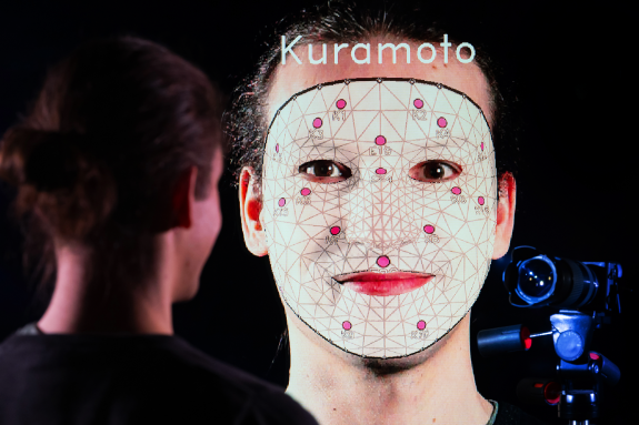
There are several classic assessment systems for classifying the severity of facial palsy. They are often subject to subjective assessments, which can vary depending on the experience of the treating physician. A data-driven approach, which uses measured facial geometry and anatomical knowledge of the facial nerve and muscles, can offer advantages here. We illustrate the current state of research in this field in an interactive demo!
Support for biodiversity research
The current climate change is leading to far-reaching changes in our flora and fauna. At the same time, a decline in biodiversity can be observed.
These trends can be measured and verified manually on a small scale in specially designed studies. On a global scale, however, this is often difficult due to the data situation.
Sensor-based systems with automatic evaluation offer an opportunity to make reliable statements on a large scale. Drones, airplanes or satellites can be used to obtain a large amount of extensive sensor data relatively quickly.
However, all of this data must then be analyzed! We show how this can be done fully automatically using the concrete example of the visual analysis of plant communities.
One aspect of this is the proportional determination of plant species on a specific area. At the same time, an assessment is made of the growth stage of plants or whether they have perhaps already died. In our exhibition area, we demonstrate such a system, which reflects the current state of research in this area.